
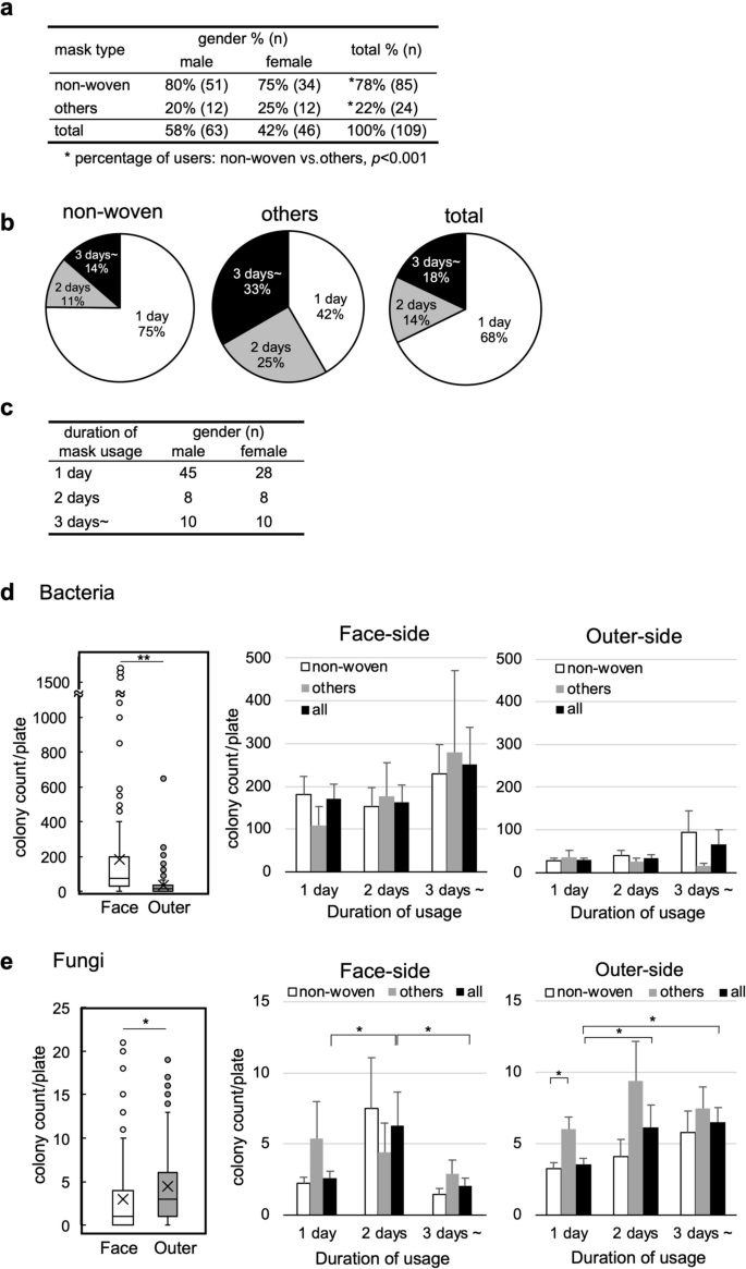
Mask types, gender differences, and duration of mask usage
Although the numbers of COVID-19 patients were relatively low in Japan during the study period, most people wore face masks in public places, and all survey participants wore face masks. First, we collected information about the mask types and duration of mask usage from 109 participants: 63 male (58%) and 46 female (42%). The majority (78% in total) of the participants used non-woven masks (Fig. 2a); the percentage of the non-woven mask users was significantly higher than that of the other mask type users (P < 0.001, most of them were polyurethane mask users except a few gauze or cloth mask users). Regarding the duration of mask usage, we found that 75% of non-woven mask users wore the masks for a single day. In contrast, 58% of the other mask type users wore the same masks for two days or more (Fig. 2b). This could be because other mask types, including polyurethane, gauze, and cloth masks, are designed washable for repeated usage; the users commonly washed and reused their masks multiple times. On the other hand, we found no significant differences between genders regarding the mask types and usage duration (Fig. 2a,c).
Microbial counts on the face-side and outer-side of masks
Microbes on the masks were cultured by pressing the face-side and outer-side of the masks onto agar plates (two plates per participant: the face-side and outer-side). We incubated the agar plates for 18 hours (h) and 5 days for bacterial and fungal propagation, respectively, and conducted colony counting.
Bacteria (Fig. 2d): We observed bacterial colonies in 99% of the samples on the face-side and 94% on the outer-side; no colony was seen in one sample on the face-side and six samples on the outer-side. The colony counts of the face-side and outer-side were 168.6 ± 24.7 and 36.0 ± 7.0 [mean ± standard error of the mean (SEM)], respectively. We compared the colony counts between the face-side and outer-side in each individual and found that the mean colony counts were 13.4-times higher on the face-side of masks (paired t-test, P < 0.001). To evaluate the influence of the mask types and duration of mask usage, we compared the colony counts among those who used the mask for one day (3–6 h), two days, and longer based on the mask types [non-woven, others, and all (non-woven and others combined)]. We found no significant differences in the colony counts among the different mask types, regardless of the duration of usage.
Fungi (Fig. 2e): We observed fungal colonies in 79% of the samples on the face-side and 95% on the outer-side. The colony counts of fungi were fewer than those of bacteria and the colony counts on the face-side and outer-side were 4.6 ± 1.9 and 6.1 ± 1.9 (mean ± SEM), respectively. In contrast to the bacterial colonies, the fungal colony counts in each individual were 2.4-times higher on the outer-side than on the face-side (paired t-test, P < 0.05). When the participants used the same masks for more than two days, the fungal colony counts were increased on the outer-side of masks, compared with the one-day usage. There were no statistical differences in the colony counts between non-woven and “others” mask users except for the fungal colony counts of the outer-side of masks after one-day usage.
Since females preferentially make up their faces, we examined whether the bacterial and fungal colony counts could be different between males and females. Only the bacterial colony counts in the face-side samples of one-day users were significantly different, lower in females (Fig. S1).
Microbial colonies and lifestyles: gargling, transportation, and natto consumption
We determined whether individual lifestyles could affect microbial counts on the masks that originate from the host (i.e., human) or the environment. One of the environmental factors that seemed to affect the levels of microbes on the masks is transportation to commute (Fig. 3a). Here, we classified into three transportation systems: (1) public transportation, including trains and buses; (2) private vehicles such as cars and trucks; and (3) walking, bicycles, and motorbikes. We found no differences in the bacterial or fungal colony counts on both sides of the masks among the three transportation systems.
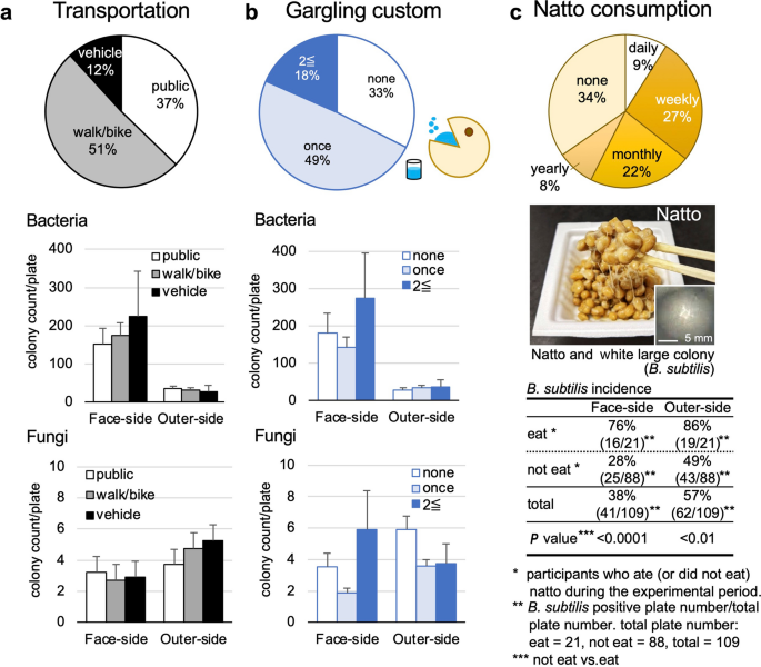
Next, we evaluated two popular habits in Japan: gargling and natto consumption. Gargling (also known as mouth/throat wash) is a Japanese custom that has been believed to prevent respiratory infections14. Of the participants, 67% gargled at least once a day and usually gargled when they returned home. However, there were no differences in the bacterial or fungal colony counts among the participants regardless of gargling (Fig. 3b).
Natto is a traditional Japanese fermented food that is sticky when eaten and clings to the mouth and chopsticks (Fig. 3c). Natto is made by fermenting soybeans with the spore-forming bacterium Bacillus subtilis, which can survive dry conditions. As expected, in this study, we observed the large white colonies formed by B. subtilis. According to the questionnaire, 9% and 27% of the participants have eaten natto daily and weekly, respectively; 19% of the participants ate natto during the experimental period. The participants who ate natto had a significantly higher incidence of large white B. subtilis colonies on both sides of the masks than those who did not.
Bacterial colony morphologies and identification
In the bacterial cultures, we observed a variety of colonies on the agar plates (Fig. 4a). We morphologically classified the colonies into four major colony forms and the other forms: (1) small white, (2) large white, (3) small yellow, (4) medium white, and the other forms, including medium to large with yellow or pink, based on the colony size (small < 2 mm, medium 2–10 mm, and large 10 mm <), color, and frequencies (Fig. 4a,b). The frequency of colonies was calculated in two formulas: (I) colony incidence = number of plates containing the colony of interest/total plate number (n = 109) × 100; and (II) % total = counts of colonies of interest/total counts of colonies in each plate × 100 (then, the mean of % total from all plates was calculated). As shown in Fig. 4a, most participants had more than one colony form. The dominance of the four colony forms regarding the colony incidence and mean % total of each colony was overall similar on the face-side and outer-side (Fig. 4b). The small white colonies were most frequently observed, with the incidence and % total exceeding 80% and 70%, respectively.
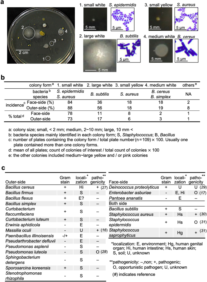
To further determine the bacteria composing each colony, we conducted Gram staining and 16S ribosomal RNA (rRNA) sequencing. The 16S rRNA sequencing showed that the small white colonies consisted mainly of Staphylococcus epidermidis, and/or S. aureus; the major bacteria species forming the small yellow colonies was S. aureus. The large white colonies were the second most observed ones and consisted of B. subtilis, a component of natto (as shown in Fig. 3c). The medium white colonies consisted of B. cereus and B. simplex; B. cereus was identified only on the outer-side of masks. Among the colonies, we also identified other bacterial species by 16S rRNA sequencing (Fig. 4c). Although most identified bacteria were non-pathogenic, there were several potential pathogenic bacteria in humans as follow: S. aureus (commensal bacterium, but its overgrowth can cause various diseases); B. cereus (intestinal bacterium, causing food poisoning); Staphylococcus saprophyticus (urinary tract infection); and Pseudomonas luteola (opportunistic pathogen)15,16,17.
Fungal colonies and identification
After quantifying fungal colonies, we further incubated them for another 2 days at 37 °C to induce spore formation. Then, using lactophenol cotton blue staining, we identified fungi on the masks based on the colony morphology macroscopically as well as the hypha and spore morphology microscopically. Although we could not identify some fungi due to lack of spore formation, we identified 13 fungal genera (Fig. 5). Among them, more than 20% of the participants had the four fungal genera, namely Cladosporium, Fonsecaea, Mucor, and Trichophyton, in common on both sides of the masks. The latter three are potentially pathogenic in humans (Fig. 5).
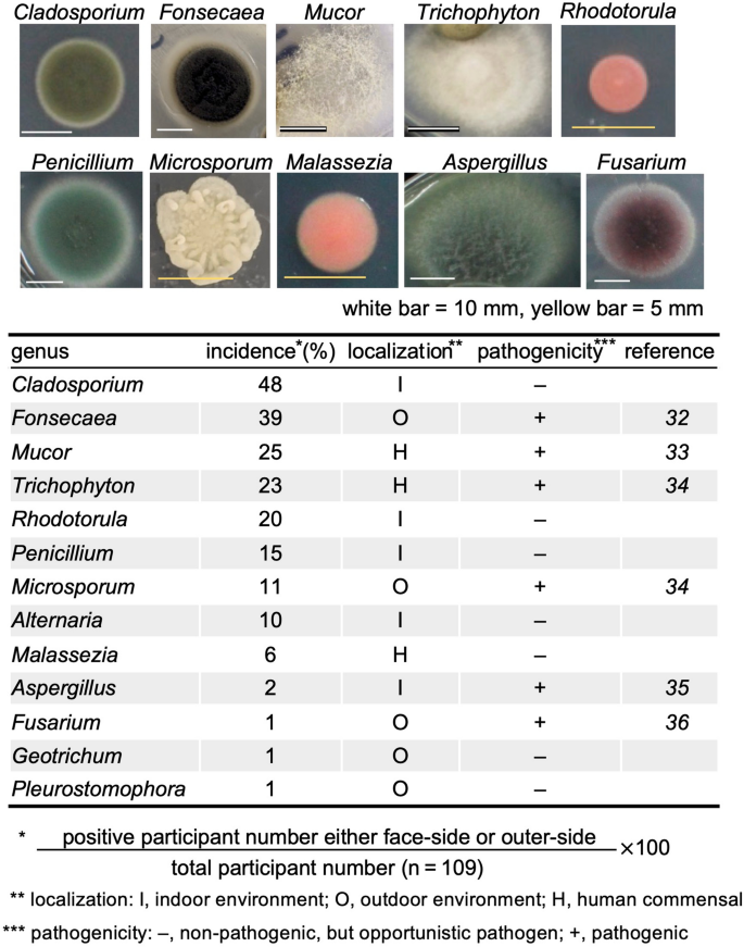
Identification of fungal colonies. We identified fungi by the colony morphology macroscopically as well as the hypha and spore morphology microscopically. Ten representative fungal images were shown. The white and yellow bars are 10 mm and 5 mm, respectively. Identified fungi, the incidence in this study, localization, and pathogenicity were listed.